Here is a collection of pictures we took over the duration of our research in the SAInT Center. Below will be brief explanations of each picture and instrument shown.
Above is a picture of the tools we used to begin our research. The following tools can be seen: Gloves, Pen, Carbon Tape, Paper Towels, 3 Different Sized Stages, 2 Different Shaped Tweezers, Scalpel, Scissors, 3D Printer Filament, Stage Holder, Dirt Container, Beaker, and Acetone. Acetone is used to clean the stages after we are done using them. Adhesive Carbon Tape is used to collect our dust samples. The Dirt Container is used to look at our dirt samples under the XRF. The Tweezers and Scalpel are used to load our samples onto the stages. The Stage and Stage Holder get loaded into the SEM when ready to take data collections. Gloves are always worn to make sure we do not contaminate the samples. The Glass Beaker is used to put the dirt samples into the oven to dry them out. A Pen is always needed to record our data. Scissors are needed to cut the 3D Filament which we analyze in the DSC and TGA. Paper Towels are used to clean up our work station after we conclude our research for the day.
As our research continued more tools were added. Above we can see a few. Plastic Wrap is needed to cover the dirt samples when placed in the XRF. This is because the XRF cannot be in direct contact with the samples, so plastic wrap is needed to make sure they do not touch. When we analyze samples that are not dirt in the XRF we use the Square Plastic Cover. It is flat and smooth and makes sure our samples do not come in contact with the XRF tip. Tape is used to label our samples, as well as, tape the plastic wrap on them to keep them from moving. A Scrapper is needed to help clean our stages and make sure the carbon tape is completely removed from the stages. Safety goggles can also be added to this list of materials. Safety goggles are mandatory and must be worn at all times when in the SAInT Center.
Above is an image of the inside of the SEM, as well as, the process of loading the SEM. Notice the "CAUTION X-RAYS" Warning Label. When handling samples gloves are always needed.
When loading the stage, the X and Y Coordinates must always be set to 20-20. This can be seen in the image above. This centers the stage so when we load and EVAC the SEM the sample will be ready to view. It also prevents the stage from hitting the top, bottom, or sides of the SEM when loading the samples. The X-Y knobs are used to move the electron beam around the sample and navigate the stage around the inside of the SEM.
Above is a picture of our main work station. This is a picture of the Scanning Electron Microscope (SEM). The monitors are used to see inside the SEM, and obtain our data.
Above is the Scanning Electron Microscope (SEM). It's a Hitachi SEM and has a Bruker attachment that will be explained later on. A SEM is a type of electron microscope that produces images of a sample by scanning it with a focused beam of electrons. The electrons interact with atoms in the sample, producing various signals that contain information about the sample's surface topography and composition. CAUTION: It releases x-rays.
Above is a picture of the Bruker Attachment that is attached to the SEM. Bruker offers a powerful range of systems of Energy-Dispersive (EDS) and Wavelength-Dispersive (WDS) X-Ray Spectrometry Electron Backscatter Diffraction Analysis (EBSD), as well as, Micro-X-Ray Flourescence and micro computed tomography on the electron microscope. It is a valuable asset to our SEM Research.
Above is the left monitor we use for the SEM. Here we use the various knobs below the screen to adjust and perfect our images. The knobs are used for the adjustment of magnification, contrast, brightness, coarse and fine focus, and X-Y alignments. These help us to operate around the sample, and find a perfect location to analyze. This is always the first step in analyzing the samples in the SEM.
The image above is of the right monitor. On this monitor is where we can analyze and quantify our data. This is where we do most of our data collection, and how we discover the elements in our samples. The image has to be loaded over from the left monitor. Once it's loaded over we can do our data analysis and collections.
Above is an image of the inside of the SEM. On the side of the SEM there is a camera to see inside the SEM to help the operator navigate around. This is a little monitor located under the bigger monitors and has to be turned on and off manually. This camera CANNOT be On when using the right monitor and collecting data. It will skew our data completely and it not good for the camera. This camera is really helpful because once the SEM is vacuum sealed there is no way of seeing into the SEM without this camera.
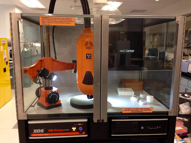
Above is a picture of the X-Ray Fluorescent (XRF) also known as the HD Prime. We use this machine along with the SEM to help us identify elements in our samples. The XRF identifies more elements than the SEM, and is beneficial for our research. A vacuum is needed over the HD Prime when operating because it releases radioactive material gases that need to be sucked out of the room to protect the operator from potentially dangerous gases. CAUTION: It releases x-rays.
Above is an image of one of the three ceiling vacuums in the SAInT Center. They suck in any airborne gases that may be released from machines or experiments that we may not be able to smell or see. This protects the researchers in the room from inhaling or in-taking any potentially dangerous gases. We placed the vacuum over the TGA and XRF when operating.
Above is a picture of the Differential Scanning Calorimeter (DSC). A DSC is a thermoanalytical technique in which difference in the amount of heat required to increase the temperature of a sample and reference is measured as a function of temperature. The DSC takes from 30 minutes to 90 minutes to complete one full measurements. This machine is different from the TGA because it is reversible, meaning the sample can be cooled, heated, and re-cooled. This means the sample can have 2 separate phase changes from solid to liquid and back to solid.
Above is the Refrigerator Cooling System that is attached to the DSC. This cools the DSC when necessary and helps our samples to complete 2 phase changes. This is beneficial because it helps us heat up and cool a sample when we need to. This is essential when we do our 3D Filament samples.
Above is a picture of a scale we use to weigh our samples and their pans. We use this scale only for when we are operating the TGA and DSC. It is very sensitive and precise with its measurements.
Above is the 3D Printer that is located in the SAInT Center. We used the filament from this printer in the DSC and TGA experiements. The 3D Printer is able to melt plastic filaments and mold them into a 3 dimensional object on its heated bed.
Above is a Thermogravimetric Analyzer (TGA). The TGA is used as a method of thermal analysis in which changes in physical and chemical properties of materials are measured as a function of increasing temperature (with constant heating rate), or as a fucntion of time (with constant temperature and/or constant mass loss). This machine takes about 45 minutes to an hour to make one successful measurement. Notice that for this machine it is imperative to use bronze tweezers over your regular aluminium or steel. Also, it is important to use the ceiling vacuums over this machine when operating. Once the phase change is complete with this machine the process is irreversible.
Above is a picture of the Oven we used to heat up and dehydrate our dirt samples. It fluctuates between 104°F and 106.5°F and we left each sample in around an hour. This is important because if any water is found in our samples when placed in the SEM it could either damage the SEM or ruin our sample.
Above is a picture of the fume hood we used when cleaning our stages. The purpose of the hood is to extract all chemical gases or fumes that may be emitted from the acetone when cleaning the stages.
Above is a picture of the DSC Pan Press. We use it to forcibly attach the pan lid to the pan with our sample inside the pan. Once the lid is on tight we can put it into the DSC and analyze the sample.
Above is a picture of an Atomic-Force Microscope (AFM). An AFM is a very high resolution type of scanning probe microscopy (SPM), with demonstrated resolution on the order of fractions of a nanometer, more than 1,000 times better than the optical diffraction limit. We have not used this machine yet by ourselves, but we plan to once we have undergone more training. We plan on doing more experiments with Graphene once we are. The first picture is the AFM itself, while the picture below it is where we conduct the experiments on the AFM. We have only ran one experiment with the AFM, and that data can be found in the Graphene blog.
*(Some definitions of the instruments were researched with the help of Wikipedia.com)*


No comments:
Post a Comment